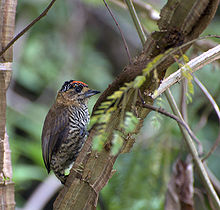
Monday 2 January 2012

""WOOD PECKER""

Woodpecker

The woodpeckers, piculets, wrynecks, and sapsuckers are a family, Picidae, of near-passerine birds. Members of this family are found worldwide, except for Australia and New Zealand, Madagascar, and the extreme polar regions. Most species live in forests or woodland habitats, although a few species are known to live in treeless areas such as rocky hillsides and deserts.
The Picidae are just one of the eight living families in the order Piciformes. Members of the order Piciformes, such as the jacamars, puffbirds, barbets, toucans, and honeyguides, have traditionally been thought to be very closely related to the woodpeckers, piculets, wrynecks, and sapsuckers. More recently, DNA sequence analyses have confirmed this view.[1]
There are about 200 species and about 30 genera in this family. Many species are threatened or endangered due to loss of habitat or habitat fragmentation. Two species of woodpeckers, the Ivory-billed Woodpecker and the Imperial Woodpecker, have been considered extinct for about 30 years (there has been some controversy recently whether these species still exist).

Contents

  • 1 General characteristics
  • 2 Distribution, habitat and movements
  • 3 Behavior
    • 3.1 Diet and feeding
    • 3.2 Breeding
  • 4 Systematics and evolution
  • 5 List of genera
  • 6 See also
  • 7 Footnotes
  • 8 References
  • 9 External links

General characteristics


The stiffened tails of woodpeckers are crucial for their climbing and foraging techniques. The tail is used as a prop. Here a Black-rumped Flameback rests while foraging using its tail for support.
The smallest woodpecker is the Bar-breasted Piculet, at 7 g and 8 cm (3¼ inches). The largest woodpecker was the Imperial Woodpecker, at an average of 58 cm (23 inches) and probably over 600 g (1.3 lb). The Ivory-billed Woodpecker is (or was) slightly smaller at 50 cm (20 inches) and a weight of 500 g (1.1 lb). If both the Ivory-billed and Imperial Woodpeckers are indeed extinct, the largest extant woodpecker is the Great Slaty Woodpecker of Southeast Asia, at about 50 cm (20 inches) and 450 g (1 lb). A number of species exhibit sexual dimorphism in size, bill length and weight. In the piculets it is often the females that are larger, amongst the woodpeckers that show sexual dimorphism it is usually the males that are larger.
Most species possess predominantly white, black, brown, green, and red plumage, although many piculets show a certain amount of grey and olive green. In woodpeckers, many species exhibit patches of red and yellow on their heads and bellies, and these bright areas are important in signaling. The dark areas of plumage are often iridescent. Although the sexes of Picidae species tend to look alike, many woodpecker species have more prominent red or yellow head markings in males than in females.
Members of the family Picidae have strong bills for drilling and drumming on trees and long sticky tongues for extracting food.[2] Woodpecker bills are typically longer, sharper and stronger than the bills of piculets and wrynecks; however their morphology is very similar. The bill's chisel-like tip is kept sharp by the pecking action in birds that regularly use it on wood. Species of woodpecker and flicker that use their bills in soil or for probing as opposed to regular hammering tend to have longer and more decurved bills. Due to their smaller bill size, many piculets and wrynecks will forage in decaying wood more often than woodpeckers. The long sticky tongues, which possess bristles, aid these birds in grabbing and extracting insects deep within a hole of a tree. It had been reported that the tongue was used to spear grubs, but more detailed studies published in 2004 have shown that the tongue instead wraps around the prey before being pulled out.[3]
Many of the foraging, breeding and signaling behaviors of woodpeckers involve drumming and hammering using the bill.[4] To prevent brain damage from the rapid and repeated decelerations, woodpeckers have evolved a number of adaptations to protect the brain. These include small brain size, the orientation of the brain within the skull (which maximises the area of contact between the brain and the skull) and the short duration of contact. The millisecond before contact with wood a thickened nictitating membrane closes, protecting the eye from flying debris.[5] The nostrils are also protected; they are often slit-like and have special feathers to cover them.
Woodpeckers, piculets and wrynecks all possess zygodactyl feet. Zygodactyl feet consist of four toes, the first (hallux) and the fourth facing backward and the second and third facing forward. This foot arrangement is good for grasping the limbs and trunks of trees. Members of this family can walk vertically up a tree trunk, which is beneficial for activities such as foraging for food or nest excavation. In addition to the strong claws and feet, woodpeckers have short strong legs. This is typical of birds that regularly forage on trunks. The tails of all woodpeckers except the piculets and wrynecks are stiffened, and when the bird perches on vertical surfaces, the tail and feet work together to support it.[2]

Distribution, habitat and movements


Use of cacti for breeding and roosting holes allows some woodpeckers, like this Ladder-backed Woodpecker, to live in otherwise treeless deserts
The woodpeckers have a mostly cosmopolitan distribution, although they are absent from Australasia, Madagascar, and Antarctica. They are also absent from the world's oceanic islands, although many insular species are found on continental islands. The true woodpeckers, subfamily Picinae, are distributed across the entire range of the woodpeckers. The Picumninae piculets have a pantropical distribution, with species in Southeast Asia, Africa, and the Neotropics, with South America holding the majority of piculet species. The second piculet subfamily, Nesoctitinae, has a single species, the Antillean Piculet, which is restricted to the Caribbean island of Hispaniola. The wrynecks (Jynginae) have an exclusively Old World distribution, with the two species occurring in Europe, Asia, and Africa.
Overall the woodpeckers are arboreal birds of wooded habitats. They reach their greatest diversity in tropical rainforests, but occur in almost all suitable habitats including woodlands, savannahs, scrublands, bamboo forests. Even grasslands and deserts have been colonised by various species. These habitats are more easily occupied where a small number of trees exist, or, in the case of desert species like the Gila Woodpecker, tall cacti are available for nesting in.[6] A number of species are adapted to spending a portion of their time feeding on the ground, and a very small minority of species have abandoned trees entirely and nest in holes in the ground. The Ground Woodpecker is one such species, inhabiting the rocky and grassy hills of South Africa
Picidae species can either be sedentary or migratory. Many species are known to stay in the same area year-round while others travel great distances from their breeding grounds to their wintering grounds. For example, the Eurasian Wryneck breeds in Europe and west Asia and migrates to the Sahel in Africa in the winter.[7]
Results from the monitoring programs of the Swiss Ornithological Institute show that the breeding populations of several forest species for which deadwood is an important habitat element (black woodpecker, great spotted woodpecker, middle spotted woodpecker, lesser spotted woodpecker, green woodpecker, three-toed woodpecker as well as crested tit, willow tit and Eurasian tree creeper) have increased in the period 1990 to 2008, although not to the same extent in all species. At the same time the white-backed woodpecker extended its range in eastern Switzerland. The Swiss National Forest Inventory shows an increase in the amount of deadwood in forests for the same period. For all the mentioned species, with the exception of green and middle spotted woodpecker, the growing availability of deadwood is likely to be the most important factor explaining this population increase.

Behavior

The woodpeckers range from highly antisocial solitary species that are aggressive to other members of their species, to species that live in groups. Group-living species tend to be communal group breeders. In addition to these species, a number of species may join mixed-species feeding flocks with other insectivorous birds, although they tend to stay at the edges of these groups. Joining these flocks allows woodpeckers to decrease anti-predator vigilance and increase their feeding rate.[8] Woodpeckers are diurnal, roosting at night inside holes. In most species the roost will become the nest during the breeding season.

[edit] Diet and feeding


Holes bored by woodpeckers feeding, Gatineau Park, Quebec

A male Black Woodpecker attending its chicks
The diet of woodpeckers consists mainly of insects and their grubs taken from living and dead trees, and other arthropods, along with fruit from live trees, nuts and sap both from live trees. Ecologically, they help to keep trees healthy by keeping them from suffering mass infestations. The family is noted for its ability to acquire wood-boring grubs using their bills for hammering, but overall the family is characterized by its dietary flexibility, with many species being both highly omnivorous and opportunistic. The insect prey most commonly taken are insects found inside tree trunks, whether they are alive or rotten wood and in crevices in bark on trees. These include beetles and their grubs, ants, termites, spiders, and caterpillars. These may be obtained either by gleaning or more famously by excavating wood. Having hammered a hole into the wood the prey is excavated by a long barbed tongue. The ability to excavate allows woodpeckers to obtain tree sap, an important source of food for some species. Most famously the sapsuckers, (genus Sphyrapicus ) feed in this fashion, but the technique is not restricted to these and others such as the Acorn Woodpecker and White-headed Woodpecker also feed in this way. It was once thought that the technique was restricted to the New World, but Old World species such as the Arabian Woodpecker and Great Spotted Woodpecker also feed in this way.[2]

Breeding

All members of the family Picidae nest in cavities. Almost every species nests in tree cavities, although in deserts some species nest inside holes in cactus and a few species nest in holes dug into the earth. Woodpeckers and piculets will excavate their own nests, but wrynecks will not. The excavated nest is usually only lined from the wood chips produced as the hole was made. Many species of woodpeckers excavate one hole per breeding season, sometimes after multiple attempts. It takes around a month to finish the job. Abandoned holes are used by other birds and mammals that are secondary cavity nesters.[9] Because nesting holes are in great demand by other cavity nesters, woodpeckers face competition for the nesting sites they excavate from the moment the hole becomes usable. This may come from other species of woodpecker, or other cavity nesting birds like swallows and starlings. Woodpeckers may aggressively harass potential competitors, and also use other strategies to reduce the chance of being usurped from their nesting site; for example the Red-crowned Woodpecker digs its nest in the underside of a small branch, which reduces the chance that a larger species will take it over and expand it.[10]
Members of Picidae are typically monogamous, with a few species breeding cooperatively and some polygamy reported in a few species.[11] Polyandry, where a female raises two broods with two separate males, has also been reported in the West Indian Woodpecker.[12] A pair will work together to help build the nest, incubate the eggs and raise their altricial young. However, in most species the male does most of the nest excavation and takes the night shift while incubating the eggs. A nest will usually consist of 2-5 round white eggs. Since these birds are cavity nesters, their eggs do not need to be camouflaged and the white color helps the parents to see them in dim light. The eggs are incubated for about 11–14 days before the chicks are born. It takes about 18–30 days before the young are ready to leave the nest.

Systematics and evolution

The phylogeny has been updated according to new knowledge about convergence patterns and evolutionary history.[13] Most notably, the relationship of the picine genera has been largely clarified, and it was determined that the Antillean Piculet is a surviving offshoot of proto-woodpeckers.
The evolutionary history of this group is not well documented, but the known fossils allow some preliminary conclusions: the earliest known modern picids were piculet-like forms of the Late Oligocene, about 25 million years ago (mya). By that time, however, the group was already present in the Americas and Europe, and it is hypothesized that they actually evolved much earlier, maybe as early as the Early Eocene (50 mya). The modern subfamilies appear to be rather young by comparison; until the mid-Miocene (10-15 mya), all picids seem to have been small or mid-sized birds similar to a mixture between a piculet and a wryneck. On the other hand, there exists a feather enclosed in fossil amber from the Dominican Republic, dated to about 25 mya, which seems to indicate that the Nesoctitinae were already a distinct lineage by then.[14]
Prehistoric representatives of the extant Picidae genera are treated in the genus articles. An enigmatic form based on a coracoid found in Pliocene deposits of New Providence, Bahamas, has been described as Bathoceleus hyphalus and probably also is a woodpecker.[15]

List of genera


Young Eurasian Wryneck (Jynx torquilla), a relative of the woodpeckers

Ochre-collared Piculet (Picumnus temminckii)

Female Yellow-shafted Flicker (Colaptes auratus auratus)
FAMILY: PICIDAE
  • Basal
    • Genus: Palaeopicus (Late Oligocene of France)
  • Incertae sedis
    • Picidae gen. et sp. indet. (Middle Miocene of New Mexico, USA)
    • Picidae gen. et sp. indet. (Late Miocene of Gargano Peninsula, Italy)
  • Subfamily: Jynginae - Wrynecks
    • Genus: Jynx (2 species)
  • Subfamily: Picumninae - Typical piculets
    • Genus: Picumnus - American Piculets (c.27 species)
    • Genus: Verreauxia - African Piculet (sometimes included in Sasia)
    • Genus: Sasia - Asian Piculets (2 species)
  • Subfamily: Nesoctitinae
    • Genus Nesoctites - Antillean Piculet
  • Subfamily: Picinae - Woodpeckers
    • Incertae sedis
      • Genus: Palaeonerpes (Ogalalla Early Pliocene of Hitchcock County, USA) - possibly dendropicine
      • Genus: Pliopicus (Early Pliocene of Kansas, USA) - possibly dendropicine
      • cf. Colaptes DMNH 1262 (Early Pliocene of Ainsworth, USA) - malarpicine?
    • Tribe: Dendropicini
      • Genus: Melanerpes (some 22 species)
      • Genus: Sphyrapicus - sapsuckers (4 species)
      • Genus: Xiphidiopicus - Cuban Green Woodpecker (Placement in Dendropicini tentative)
      • Genus: Dendropicos (15 species)
      • Genus: Mesopicos (3 species)
      • Genus: Dendrocopos (21 species)
      • Genus: Picoides (presently 12 species; maybe only 3 belong here) - this genus is in need of revision.[16] See the genus article for more.
      • Genus: Veniliornis (14 species)
    • Tribe: Malarpicini
      • Genus: Campethera (12 species)
      • Genus: Geocolaptes - Ground Woodpecker
      • Genus: Dinopium - malarpicine flamebacks (4 species)
      • Genus: Meiglyptes (3 species)
      • Genus: Hemicircus (2 species; placement in Malarpicini tentative)
      • Genus: Micropternus - Rufous Woodpecker (formerly in Celeus)
    • Tribe: Picini (sometimes included in Malarpicini)
      • Genus: Picus (c. 15 species)
      • Genus: Mulleripicus (3 species)
      • Genus: Dryocopus (7 species)
      • Genus: Celeus (11 species)
      • Genus: Piculus (7 species)
      • Genus: Colaptes - flickers (about 12 species)
    • Tribe: Megapicini
      • Genus: Campephilus (11 species, 2 possibly recently extinct)
      • Genus: Chrysocolaptes - megapicine flamebacks (2 species)
      • Genus: Reinwardtipicus - Orange-backed Woodpecker
      • Genus: Blythipicus (2 species)
      • Genus: Gecinulus (2 species; placement in Megapicini tentative)
      • Genus: Sapheopipo - Okinawa Woodpecker (Placement in Megapicini tentative)

STAR FRUIT TREE

Carambola


Carambolas still on the tree
Carambola, also known as starfruit, is the fruit of Averrhoa carambola, a species of tree native to the Philippines, Indonesia, Malaysia, India, Bangladesh, and Sri Lanka. The fruit is a popular food throughout Southeast Asia, the South Pacific and parts of East Asia. The tree is also cultivated throughout non-indigenous tropical areas, such as in Costa Rica, Peru, Colombia, Jamaica, Trinidad, Ecuador, Guyana, Dominican Republic, Puerto Rico, Haiti, and Brazil, and, in the United States, in south Florida and Hawaii.
The fruit has ridges running down its sides (usually five); in cross-section, it resembles a star, hence its name. The number of ridges can vary from three to six.

Contents

  • 1 Origins and distribution
  • 2 Nomenclature
  • 3 Gastronomy
  • 4 Health
    • 4.1 Benefits
    • 4.2 Risks
    • 4.3 Drug interactions
  • 5 Cultivation
  • 6 References

Origins and distribution

The carambola has been cultivated in parts of Asia for hundreds of years. Scientists believe that it may have originated in Sri Lanka or Moluccas, Indonesia.
Due to concerns over pests and pathogens, however, whole starfruits cannot yet be imported to the US from Malaysia under current Food and Drug Administration regulations. In the United States, starfruits are grown in tropical and semitropical areas, including Florida, Puerto Rico, and Hawaii.

  Nomenclature

The carambola is known under different names in different countries. It should not be confused with the closely related bilimbi, with which it shares some common names.
  • Assamese - kordoi (কৰ্দৈ) / rohdoi (ৰহদৈ)
  • Bengali - kamranga
  • English - carambola, starfruit
  • Filipino - balimbing, saranate
  • Hindi - kamrakh
  • Tamil - thambaratham (தம்பரத்தம்)
  • Indonesian - belimbing
  • Vietnamese - khế

  Gastronomy


Vertical, side and cross section profiles of ripe carambolas
Carambola, raw
Nutritional value per 100 g (3.5 oz)
Energy 128 kJ (31 kcal)
Carbohydrates 6.73 g
- Sugars 3.98 g
- Dietary fiber 2.8 g
Fat .33 g
Protein 1.04 g
Pantothenic acid (B5) .39 mg (8%)
Folate (vit. B9) 12 μg (3%)
Vitamin C 34.4 mg (41%)
Phosphorus 12 mg (2%)
Potassium 133 mg (3%)
Zinc .12 mg (1%)
Percentages are relative to US recommendations for adults.
Source: USDA Nutrient Database
The entire fruit is edible, including the slightly waxy skin, unlike other tropical fruits. The flesh is crunchy, firm, and extremely juicy, having a texture similar in consistency to that of grapes.
Carambolas are best consumed when ripe, when they are yellow with a light shade of green. They will also have brown ridges at the five edges and feel firm. Overripe starfruit will be yellow with brown spots and can become soggier in consistency.
Ripe carambolas are sweet without being overwhelming, and have a tart, sour undertone. The taste is difficult to compare, but it has been likened to a mix of apple, pear, and citrus family fruits all at once. Unripe starfruits are firmer and sour, and taste like green apples.

HealthBenefits


Carambolas in varying stages of ripeness
Carambola is rich in antioxidants and vitamin C and low in sugar, sodium, and acid. It is also a potent source of both primary and secondary polyphenolic antioxidants.A. carambola has both antioxidant and antimicrobial activities: Scavenging of NO by the fruit extract is dependent on concentration and stage of ripening. Extracts showed antimicrobial activity against E. coli, Salmonella typhi, Staphylococcus aureus, and Bacillus cereus.

Risks

Carambola is a fairly complex fruit with many benefits, but, as with strawberries[citation needed], a small percentage of the human population should be cautious of the fruit for health reasons. Carambola contains oxalic acid, which can be harmful to individuals suffering from kidney failure, kidney stones, or those under kidney dialysis treatment. Consumption by those with kidney failure can produce hiccups, vomiting, nausea, and mental confusion. Fatal outcomes have been documented in some patients.

Drug interactions

Like the grapefruit, carambola is considered to be a potent inhibitor of seven cytochrome P450 isoforms. These enzymes are significant in the first-pass elimination of many medicines, and, thus, the consumption of carambola or its juice in combination with certain medications can significantly increase their effective dosage within the body. Research into grapefruit juice has identified a number of common medications affected, including statins, which are commonly used to treat cardiovascular illness, and benzodiazepines (a tranquilizer family including diazepam).

cultivation

Ripening carambolas still on the tree
The carambola is a tropical and subtropical fruit. It can be grown at up to 4,000 feet (1,200 m) in elevation. It prefers full sun exposure, but requires enough humidity and a total of 70 inches or more of rainfall a year. It does not have a soil type preference, but it requires good drainage.
Carambola trees are planted at least 20 feet (6.1 m) from each other and typically are fertilized three times a year. The tree grows rapidly and typically produces fruit at four or five years of age. The large amount of rain during spring actually reduces the amount of fruit, but, in ideal conditions, carambola can produce from 200 to 400 pounds (91 to 180 kg) of fruit a year. The fruit is harvested mainly during the months of June, July, and August, but sometimes year-round.
Major pests are fruit flies, ants, and birds. Crops are also susceptible to frosts, especially in the United States and in the Philippines.
Malaysia is the global leader in starfruit production by volume and ships the product all over Asia and Europe.

""STAR FISH""

Starfish

Sea Stars
Temporal range: Ordovician–Recent
PreЄ
Є
O
S
D
C
P
T
J
K
Pg
N

"Asteroidea" from Ernst Heckel's
Scientific classification
Kingdom: Animalia
Phylum: Echinodermata
Subphylum: Asterozoa
Class: Asteroidea
De Blainville, 1830
Orders
Brisingida (100 species)
Forcipulatida (300 species)
Paxillosida (255 species)
Notomyotida (75 species)
Spinulosida (120 species)
Valvatida (695 species)
Velatida (203 species)
Starfish or sea stars are echinoderms belonging to the class Asteroidea. The names "starfish" and "sea star" essentially refer to members of the class Asteroidea. However, common usage frequently finds "starfish" and "sea star" also applied to ophiuroids which are correctly referred to as "brittle stars" or "basket stars".
There are about 1,800 living species of starfish that occur in all the world's oceans, including the Atlantic, Pacific, Indian as well as in the Arctic and the Southern Ocean (i.e., Antarctic) regions. Starfish occur across a broad depth range from the intertidal to abyssal depths (>6000 m).
Starfish are among the most familiar of marine animals and possess a number of widely known traits, such as regeneration and feeding on mussels. Starfish possess a wide diversity of body forms and feeding methods. The extent that Asteroidea can regenerate varies with individual species. Broadly speaking, starfish are opportunistic feeders, with several species having specialized feeding behavior, including suspension feeding and specialized predation on specific prey.
The Asteroidea occupy several important roles throughout ecology and biology. Sea stars, such as the Ochre sea star (Pisaster ochraceus) have become widely known as the example of the keystone species concept in ecology. The tropical Crown of Thorns starfish (Acanthaster planci) are voracious predators of coral throughout the Indo-Pacific region. Other starfish, such as members of the Asterinidae, are frequently used in developmental biology.

Contents

  • 1 Appearance
  • 2 Internal anatomy
    • 2.1 Digestive system
    • 2.2 Nervous system
    • 2.3 Locomotion
    • 2.4 Endoskeleton
    • 2.5 Respiration and excretion
  • 3 Life cycle
    • 3.1 Sexual reproduction
    • 3.2 Asexual reproduction
    • 3.3 Larval development
    • 3.4 Lifespan
    • 3.5 Regeneration
  • 4 Diet
  • 5 Distribution
  • 6 Diversity
  • 7 Threats
  • 8 References
  • 9 Further reading
  • 10 External links
  • 11 Gallery

Appearance


Red-knobbed starfish Protoreaster linckii, a sea star from the Indian Ocean

Schmedelian pin-cushion sea star (Culcita schmideliana) on Meedhupparu house reef in the Maldives

Closeup of the top surface of a starfish
Starfish express pentamerism or pentaradial symmetry as adults.[clarification needed] However, theoretically, the evolutionary ancestors of echinoderms are believed to have had bilateral symmetry. Starfish, as well as other echinoderms, do exhibit bilateral symmetry, but only as larval forms.
Most starfish typically have five rays or arms, which radiate from a central disk. However, several species frequently have six or more arms. Several asteroid groups, such as the Solasteridae, have 10–15 arms whereas some species, such as the Antarctic Labidiaster annulatus can have up to 50. It is not unusual for species that typically have five-rays to exceptionally possess six or more rays due to developmental abnormalities.
The bodies of starfish are composed of calcium carbonate components, known as ossicles. These form the endoskeleton, which takes on a variety of forms that are externally expressed as a variety of structures, such as spines and granules. The architecture and individual shape/form of these plates which often occur in specific patterns or series, as well as their location are the source of morphological data used to classify the different groups within the Asteroidea. Terminology referring to body location in sea stars is usually based in reference to the mouth to avoid incorrect assumptions of homology with the dorsal and ventral surfaces in other bilateral animals. The bottom surface is often referred to as the oral or actinal surface whereas the top surface is referred to as the aboral or abactinal side.
The body surface of sea stars often has several structures that comprise the basic anatomy of the animal and can sometimes assist in its identification.
The madreporite can be easily identified as the light-colored circle, located slightly off center on the central disk. This is a porous plate which is connected via a calcified channel to the animal's water vascular system in the disk. Its function is, at least in part, to provide additional water for the animal's needs, including replenishing water to the water vascular system.
Several groups of asteroids, including the Valvatacea but especially the Forcipulatacea possess small bear-trap or valve-like structures known as pedicellariae. These can occur widely over the body surface. In forcipulate asteroids, such as Asterias or Pisaster, pedicellariae occur in pom-pom like tufts at the base of each spine, whereas in goniasterids, such as Hippasteria, pedicellariae are scattered over the body surface. Although the full range of function for these structures is unknown, some are thought to act as defense where others have been observed to aid in feeding. The Antarctic Labidiaster annulatus uses its large, pedicellariae to capture active krill prey. The North Pacific Stylasterias has been observed to capture small fish with its pedicellariae.
Other types of structures vary by taxon. For example, Porcellanasteridae employ additional cribriform organs which occur among their lateral plate series, which are thought to generate current in the burrows made by these infaunal sea star.

 Internal anatomy


Dissection of Asterias rubens
1 – Pyloric stomach 2 – Intestine and anus 3 – Rectal sac 4 – Stone canal 5 – Madreporite 6 – Pyloric caecum 7 – Digestive glands 8 – Cardiac stomach 9 – Gonad 10 – Radial canal 11 – Tube feet
As echinoderms, starfish possess a hydraulic water vascular system that aids in locomotion.The water vascular system has many projections called tube feet on the ventral face of the sea star's arms which function in locomotion and aid with feeding. Tube feet emerge through openings in the endoskeleton and are externally expressed through the open grooves present along the bottom of each arm.
The body cavity not only contains the water vascular system that operates the tube feet, but also the circulatory system, called the hemal system. Hemal channels form rings around the mouth (the oral hemal ring), closer to the top of the sea star and around the digestive system (the gastric hemal ring). A portion of the body cavity called the axial sinus connects the three rings. Each ray also has hemal channels running next to the gonads.
On the end of each arm or ray there is a microscopic eye (ocellus), which allows the sea star to see, although it only allows it to see light and dark, which is useful to see movement. Only part of the cells are pigmented (thus a red or black color) and there is no cornea or iris. This eye is known as a pigment spot ocellus.
Several types of toxins and secondary metabolites have been extracted from several species of sea star. Research into the efficacy of these compounds for possible pharmacological or industrial use occurs worldwide.

Digestive system

The mouth of a starfish is located on the underside of the body, and opens through a short esophagus into firstly a cardiac stomach, and then, a second, pyloric stomach. Each arm also contains two pyloric caeca, long hollow tubes branching outwards from the pyloric stomach. Each pyloric caecum is lined by a series of digestive glands, which secrete digestive enzymes and absorb nutrients from the food. A short intestine runs from the upper surface of the pyloric stomach to open at an anus in the center of the upper body.
Many sea stars, such as Astropecten and Luidia swallow their prey whole, and start to digest it in the stomachs before passing it into the pyloric caeca. However, in a great many species, the cardiac stomach can be everted out of the organism's body to engulf and digest food. In these species, the cardiac stomach fetches the prey then passes it to the pyloric stomach, which always remains internal.
Some species are able to use their water vascular systems to force open the shells of bivalve molluscs such as clams and mussels by injecting their stomachs into the shells. With the stomach inserted inside the shell, the sea star is able to digest the mollusc in place. The cardiac stomach is then brought back inside the body, and the partially digested food is moved to the pyloric stomach. Further digestion occurs in the intestine. Waste is excreted through the anus on the aboral side of the body.
Because of this ability to digest food outside of its body, the sea star is able to hunt prey that are much larger than its mouth would otherwise allow, such as clams and oysters, arthropods, small fish, and molluscs. However, some species are not pure carnivores, and may supplement their diet with algae or organic detritus. Some of these species are grazers, but others trap food particles from the water in sticky mucus strands that can be swept towards the mouth along ciliated grooves.
Some echinoderms can live for several weeks without food under artificial conditions. Scientists believe that they may receive some nutrients from organic material dissolved in seawater.

Nervous system

Echinoderms have rather complex nervous systems, with a distributed brain. All echinoderms have a network of interlacing nerves called a nerve plexus which lies within, as well as below, the skin.The esophagus is also surrounded by a central nerve ring which sends radial nerves into each of the arms, often parallel with the branches of the water vascular system. These all connect to form a brain. The ring nerves and radial nerves coordinate the sea star's balance and directional systems.[citation needed]
Although the echinoderms do not have many well-defined sensory inputs, they are sensitive to touch, light, temperature, orientation, and the status of water around them. The tube feet, spines, and pedicellariae found on sea stars are sensitive to touch, while eyespots on the ends of the rays are light-sensitive.The tube feet, especially those at the tips of the rays, are also sensitive to chemicals and this sensitivity is used in locating odor sources, such as food.
The eyespots each consist of a mass of ocelli, consisting of pigmented epithelial cells that respond to light and narrow sensory cells lying between them. Each ocellus is covered by a thick, transparent, cuticle that both protects them and acts as a lens. Many starfish also possess individual photoreceptor cells across their body and are able to respond to light even when their eyespots are covered.

Locomotion


The underside of a sea star. The inset shows a magnified view of the tube feet.
Sea stars move using a water vascular system. Water comes into the system via the madreporite. It is then circulated from the stone canal to the ring canal and into the radial canals. The radial canals carry water to the ampulla portion of tube feet.
Tube feet consist of internal ampulla and external podium, or "foot". The ampulla squeezes forcing water into podium, which expands to contact substrate. Although the podium resembles a suction cup, gripping action is a function of adhesive chemicals rather than suction. De-adhesive chemicals and podial contraction allow for the release off of substrate.
The tube feet latch on to surfaces and move in a wave, with one body section attaching to the surfaces as another releases. Most sea stars cannot move quickly. However, some burrowing species from the genera Astropecten and Luidia are capable of rapid, creeping motion: "gliding" across the ocean floor[citation needed]. This motion results from their pointed tubefeet adapted specially for excavating patches of sand[citation needed].

Sea-star endoskeleton

endoskeleton

Sea stars, like other echinoderms have mesodermal endoskeletons consisting of small calcareous ossicles (bony plates).

Respiration and excretion

Respiration occurs mainly through the tube feet, and through tiny structures called papillae that dot the body surface. These papillae are thin-walled projections of the body cavity, reaching through the muscular body wall and into the surrounding water. Oxygen from the water is distributed through the body mainly by the fluid in the main body cavity; the hemal system may also play a minor role.
Excretion of nitrogenous waste is also performed through the tube feet and papillae, and there are no distinct excretory organs. The body fluid contains phagocytic cells called coelomocytes, which are also found within the hemal and water vascular systems. These cells engulf waste material, and eventually migrate to the tips of the papillae where they are ejected into the surrounding water. Some waste may also be excreted by the pyloric glands and voided with the faeces.
Starfish do not appear to have any mechanisms for osmoregulation, and keep their body fluids at the same salt concentration as the surrounding water. Although some species can tolerate relatively low salinity, the lack of osmoregulation likely explains why starfish are not found in fresh water, or even in estuarine environments.

Life cycle

Starfish are capable of both sexual and asexual reproduction.

  Sexual reproduction


An eleven-armed sea star
Most species are dioecious, with separate male and female individuals (also referred to as gonochoric). Male and female sea stars are usually not distinguishable from the outside; one needs to see the gonads or be lucky enough to catch them spawning. Some species are simultaneous hermaphrodites (producing eggs and sperm at the same time). In a few of these the same gonad (called an ovotestes) produces eggs and sperm. Yet other sea stars are sequential hermaphrodites with some species being protandrous i.e. young individuals are males that change into females as they grow larger. (e.g. Asterina gibbosa ) and others protogynous. In some of these species, when a large female divides, the smaller individuals produced become males. When they grow big enough they change back into females.
Each arm contains two gonads, which release gametes through openings called gonoducts, located on the central body between the arms.
Fertilization is external in most species though a few show internal fertilization. In most species the buoyant eggs and sperm are simply released into the water (free spawning) and the resulting embryos and larvae live as part of the plankton. In others the eggs may be stuck onto the underside of rocks to develop.Certain species of sea star brood their eggs – either by simply sitting on them or by holding them in specialised structures. These structures include chambers on their aboral surface, the pyloric stomach(Leptasterias tenera) or even the gonads themselves.Those sea stars that brood their eggs by sitting on them usually raise their disc and assume a humped posture.One species broods a few of its young and broadcasts the remaining eggs which wont fit into the pouch. In these brooding species, the eggs are relatively large, and supplied with yolk, and they generally (but not always) develop directly into miniature starfish, without a larval stage. The developing young are called lecithotrophic because they get their nutrition from the yolk as opposed to planktotrophic feeding larvae. In one species of intragonadal brooder the young starfish get their nutrition by eating the eggs and embryos with them in their gonadal brood pouch. Brooding is especially common in polar and deep-sea species, environments less favourable for larvae and in smaller species that produce few eggs.
To increase their chances of fertilization, sea stars may synchronize their spawning, gather in group or form pairs. This is called pseudo-copulation when the males climb onto the females with their arms placed between those of the female and release the sperm around them. Sea stars may use environmental signals to coordinate timing of spawning (day length to indicate the correct time of the year, dawn or dusk to indicate the correct time of day), and chemical signals to indicate their readiness to each other.In some species mature females produce chemicals that attract sperm in the sea water.

 Asexual reproduction

Some species of sea star also reproduce asexually by fragmentation, often with part of an arm becoming detached and eventually developing into an independent individual sea star. Sea stars can be pests to fishermen who make their living on the capture of clams and other mollusks at sea as sea stars prey on these. The fishermen would think they had killed the sea stars by chopping them up and disposing of them at sea, but each fragment would regenerate into a complete adult, ultimately leading to their increased numbers until the issue was better understood[citation needed]. A sea-star arm can even regenerate into a whole new organism if none of the central disk of the sea star is part of the chopped off arm. Even the tip of an arm can regenerate into a new individual. A starfish which is regenerating from a severed arm, with one full-sized arm and the other arms small, is sometimes called a "comet starfish".

Larval development

Like all echinoderms, starfish are developmentally (embryologically) deuterostomes; a feature they share with chordates (including vertebrates), but not with most other invertebrates. Their embryo initially develops bilateral symmetry, again reflecting their likely common ancestry with chordates. Later development takes a very different path, however, as the developing star fish settles out of the zooplankton and develops the characteristic radial symmetry. As the organism grows, one side of the body grows more than the other, and eventually absorbs the smaller side. After that, the body is formed into five parts around a central axis. Then the echinoderm has radial symmetry.
The larvae of echinoderms are ciliated, free-swimming organisms. Fertilized eggs grow into bipinnaria and (in most cases) later into brachiolaria larvae, which either grow using a yolk or by catching and eating other plankton. In either case, they live as plankton, suspended in the water and swimming by using beating cilia. The larvae are bilaterally symmetric — unlike adults, they have a distinct left and right side. Eventually, they undergo a complete metamorphosis, settle to the bottom, and grow into adults.

Lifespan

The lifespan of starfish varies considerably between species, generally being longer in larger species. For example, Leptasterias hexactis (adult weight 2 grams) reaches sexual maturity in two years, and lives for about ten years in total, while Pisaster ochraceus (adult weight 80 grams) reaches maturity in five years, and may live to the age of 34.

Regeneration


A sunflower starfish is regenerating arms that were lost to predators
Some species of sea star have the ability to regenerate lost arms and can regrow an entire new arm in time. Sea stars can grow an entire sea star from a single ray, as in the red and blue Linckia star.

Diet


Sea star Pisaster ochraceus consuming a mussel in Central California
Most species are generalist predators, eating mollusks such as clams, oysters, some snails, or any other animal too slow to evade the attack (e.g. other echinoderms, or dying fish). Some species are detritivores, eating decomposed animal and plant material or organic films attached to substrate. Others may consume coral polyps (the best-known example for this is the infamous Crown-of-thorns starfish), sponges or even suspended particles and plankton (such as sea stars of the Order Brisingida).The processes of feeding and capture may be aided by special parts; Pisaster brevispinus or short-spined pisaster from the West Coast of America may use a set of specialized tube feet to extend itself deep into the soft substrata to extract prey (usually clams). Grasping the shellfish, the sea star slowly pries open the shell by wearing out the adductor muscle and then inserts (also called evisceration) its stomach into an opening to devour the organism.

Distribution

There are about 1,800 known living species of sea star, and they occur in all of the Earth's oceans. The greatest variety of sea stars is found in the tropical Indo-Pacific. Areas known for their great diversity include the tropical-temperate regions around Australia, the tropical East Pacific, and the cold-temperate water of the North Pacific (California to Alaska). Asterias is a common genus found in European waters and on the eastern coast of the United States; Pisaster, along with Dermasterias ("leather star"), are usually found on the western coast. Habitats range from tropical coral reefs, kelp forests to deep-sea floor, although none of them live within the water column; all species of sea star found are living as benthos. Echinoderms need a delicate internal balance in their body; no sea stars are found in freshwater environments.

 Diversity


Sea stars move using a water vascular system. Water comes into the system via the madreporite.
As mentioned above, there are over 1,800 species; with many species awaiting discovery. Some of the better known sea stars would include:
  • Bat star
  • Blue sea star
  • Carpet sea star
  • Comb sea star
  • Common starfish
  • Crown-of-thorns sea star
  • Eleven-armed sea star
  • Japanese sea star
  • Ochre sea star
  • Pincushion sea star
  • Pink sea star
  • Horned sea star
The Northern Pacific sea star (Asterias amurensis) known as gohongaze is considered an edible delicacy.[

 Threats

Sea stars and other echinoderms pump water directly into their bodies, via the water vascular system, as they find it. This makes them vulnerable to all forms of water pollution, as they have little ability to filter the water of toxins and contaminants. Oil spills and similar events often take a toll on echinoderm populations that carry consequences for the ecosystem.

STOMACH CANCER

Stomach cancer

Gastric cancer
Classification and external resources

A suspicious stomach ulcer that was diagnosed as cancer on biopsy and resected. Surgical specimen.
ICD-10 C16
ICD-9 151.9
OMIM 137215
DiseasesDB 12445
eMedicine med/845
MeSH D013274
Gastric cancer, commonly referred to as stomach cancer, can develop in any part of the stomach and may spread throughout the stomach and to other organs; particularly the esophagus, lungs, lymph nodes, and the liver. Stomach cancer causes about 800,000 deaths worldwide per year.

Contents

  • 1 Signs and symptoms
  • 2 Causes
  • 3 Diagnosis
    • 3.1 Histopathology
    • 3.2 Staging
  • 4 Management
    • 4.1 Surgery
    • 4.2 Chemotherapy
    • 4.3 Radiation
    • 4.4 Multimodality therapy
  • 5 Epidemiology
  • 6 In other animals
  • 7 References
  • 8 External links

Signs and symptoms


Endoscopic image of linitis plastica, a type of stomach cancer where the entire stomach is invaded, leading to a leather bottle-like appearance with blood coming out of it.

Endoscopic image of early stage of the stomach cancer. Its histology was poorly differentiated adno carcinoma with signet ring cells.Left above=Normal, right above=FICE, left low=acetate stained, right low= AIM stained
Stomach cancer is often asymptomatic or causes only nonspecific symptoms in its early stages. By the time symptoms occur, the cancer has often reached an advanced stage (see below), one of the main reasons for its poor prognosis.[citation needed] Stomach cancer can cause the following signs and symptoms:
Stage 1 (Early)
  • Indigestion or a burning sensation (heartburn)
  • Loss of appetite, especially for meat
  • Abdominal discomfort or irritation
Stage 2 (Middle)
  • Weakness and fatigue
  • Bloating of the stomach, usually after meals
Stage 3 (Late)
  • Abdominal pain in the upper abdomen
  • Nausea and occasional vomiting
  • Diarrhea or constipation
  • Weight loss
  • Bleeding (vomiting blood or having blood in the stool) which will appear as black. This can lead to anemia.
  • Dysphagia; this feature suggests a tumor in the cardia or extension of the gastric tumor in to the esophagus.
Note that these can be symptoms of other problems such as a stomach virus, gastric ulcer or tropical sprue.

 Causes

Infection by Helicobacter pylori is believed to be the cause of most stomach cancer while autoimmune atrophic gastritis, intestinal metaplasia and various genetic factors are associated with increased risk levels. The Merck Manual states that diet plays no role in the genesis of stomach cancer. However, the American Cancer Society lists the following dietary risks, and protective factors, for stomach cancer: "smoked foods, salted fish and meat, and pickled vegetables (appear to increase the risk of stomach cancer.) Nitrates and nitrites are substances commonly found in cured meats. They can be converted by certain bacteria, such as H. pylori, into compounds that have been found to cause stomach cancer in animals. On the other hand, eating fresh fruits and vegetables that contain antioxidant vitamins (such as A and C) appears to lower the risk of stomach cancer." A December 2009 article in American Journal of Clinical Nutrition found a statistically significant inverse correlation between higher adherence to a Mediterranean Dietary Pattern and stomach cancer.
In more detail, H. pylori is the main risk factor in 65–80% of gastric cancers, but in only 2% of such infections. Approximately ten percent of cases show a genetic component. Some studies indicate that bracken consumption and spores are correlated with incidence of stomach cancer, though causality has yet to be established.
A very important but preventable cause of gastric cancer is tobacco smoking. Smoking increases the risk of developing gastric cancer considerably; from 40% increased risk for current smokers to 82% increase for heavy smokers which is nearly twice the risk for non-smoking population. Gastric cancers due to smoking mostly occur in upper part of stomach near esophagusAnother lifestyle cause of gastric cancer besides smoking is consumption of alcohol.Alcohol as cause of cancer along with tobacco smoking as cause of cancer increase the risk of developing other cancers as well.
Gastric cancer shows a male predominance in its incidence as up to three males are affected for every female. Estrogen may protect women against the development of this cancer form.A very small percentage of diffuse-type gastric cancers (see Histopathology below) are thought to be genetic. Hereditary Diffuse Gastric Cancer (HDGC) has recently been identified and research is ongoing. However, genetic testing and treatment options are already available for families at risk.
Some researchers showed a correlation between Iodine deficiency or excess, iodine-deficient goitre and gastric cancer; a decrease of the incidence of death rate from stomach cancer after implementation of the effective I-prophylaxis was reported too. The proposed mechanism of action is that iodide ion can function in gastric mucosa as an antioxidant reducing species that can detoxify poisonous reactive oxygen species, such as hydrogen peroxide.
The International Cancer Genome Consortium is leading efforts to map stomach cancer's complete genome.[citation needed]

Diagnosis

To find the cause of symptoms, the doctor asks about the patient's medical history, does a physical exam, and may order laboratory studies. The patient may also have one or all of the following exams:
  • Gastroscopic exam is the diagnostic method of choice. This involves insertion of a fiber optic camera into the stomach to visualize it.
  • Upper GI series (may be called barium roentgenogram)
  • Computed tomography or CT scanning of the abdomen may reveal gastric cancer, but is more useful to determine invasion into adjacent tissues, or the presence of spread to local lymph nodes.
Abnormal tissue seen in a gastroscope examination will be biopsied by the surgeon or gastroenterologist. This tissue is then sent to a pathologist for histological examination under a microscope to check for the presence of cancerous cells. A biopsy, with subsequent histological analysis, is the only sure way to confirm the presence of cancer cells.
Various gastroscopic modalities have been developed to increased yield of detect mucosa with a dye that accentuates the cell structure and can identify areas of dysplasia. Endocytoscopy involves ultra-high magnification to visualize cellular structure to better determine areas of dysplasia. Other gastroscopic modalities such as optical coherence tomography are also being tested investigationally for similar applications.
A number of cutaneous conditions are associated with gastric cancer. A condition of darkened hyperplasia of the skin, frequently of the axilla and groin, known as acanthosis nigricans, is associated with intra-abdominal cancers such as gastric cancer. Other cutaneous manifestations of gastric cancer include tripe palms (a similar darkening hyperplasia of the skin of the palms) and the sign of Leser-Trelat, which is the rapid development of skin lesions known as seborrheic keratoses.
Various blood tests may be done; including: Complete Blood Count (CBC) to check for anemia. Also, a stool test may be performed to check for blood in the stool.

 Histopathology


Poor to moderately differentiated adenocarcinoma of the stomach. H&E stain.

Gastric signet ring cell carcinoma. H&E stain.

Adenocarcinoma of the stomach and intestinal metaplasia. H&E stain.
  • Gastric adenocarcinoma is a malignant epithelial tumor, originating from glandular epithelium of the gastric mucosa. Stomach cancers are overwhelmingly adenocarcinomas (90%). Histologically, there are two major types of gastric adenocarcinoma (Lauren classification): intestinal type or diffuse type. Adenocarcinomas tend to aggressively invade the gastric wall, infiltrating the muscularis mucosae, the submucosa, and thence the muscularis propria. Intestinal type adenocarcinoma tumor cells describe irregular tubular structures, harboring pluristratification, multiple lumens, reduced stroma ("back to back" aspect). Often, it associates intestinal metaplasia in neighboring mucosa. Depending on glandular architecture, cellular pleomorphism and mucosecretion, adenocarcinoma may present 3 degrees of differentiation: well, moderate and poorly differentiated. Diffuse type adenocarcinoma (mucinous, colloid, linitis plastica, leather-bottle stomach) Tumor cells are discohesive and secrete mucus which is delivered in the interstitium producing large pools of mucus/colloid (optically "empty" spaces). It is poorly differentiated. If the mucus remains inside the tumor cell, it pushes the nucleus to the periphery- "signet-ring cell".
  • Around 5% of gastric malignancies are lymphomas (MALTomas, or MALT lymphoma).
  • Carcinoid and stromal tumors may also occur.

Staging

If cancer cells are found in the tissue sample, the next step is to stage, or find out the extent of the disease. Various tests determine whether the cancer has spread and, if so, what parts of the body are affected. Because stomach cancer can spread to the liver, the pancreas, and other organs near the stomach as well as to the lungs, the doctor may order a CT scan, a PET scan, an endoscopic ultrasound exam, or other tests to check these areas. Blood tests for tumor markers, such as carcinoembryonic antigen (CEA) and carbohydrate antigen (CA) may be ordered, as their levels correlate to extent of metastasis, especially to the liver, and the cure rate.
Staging may not be complete until after surgery. The surgeon removes nearby lymph nodes and possibly samples of tissue from other areas in the abdomen for examination by a pathologist.
The clinical stages of stomach cancer are:
  • Stage 0. Limited to the inner lining of the stomach. Treatable by endoscopic mucosal resection when found very early (in routine screenings); otherwise by gastrectomy and lymphadenectomy without need for chemotherapy or radiation.
  • Stage I. Penetration to the second or third layers of the stomach (Stage 1A) or to the second layer and nearby lymph nodes (Stage 1B). Stage 1A is treated by surgery, including removal of the omentum. Stage 1B may be treated with chemotherapy (5-fluorouracil) and radiation therapy.
  • Stage II. Penetration to the second layer and more distant lymph nodes, or the third layer and only nearby lymph nodes, or all four layers but not the lymph nodes. Treated as for Stage I, sometimes with additional neoadjuvant chemotherapy.
  • Stage III. Penetration to the third layer and more distant lymph nodes, or penetration to the fourth layer and either nearby tissues or nearby or more distant lymph nodes. Treated as for Stage II; a cure is still possible in some cases.
  • Stage IV. Cancer has spread to nearby tissues and more distant lymph nodes, or has metastatized to other organs. A cure is very rarely possible at this stage. Some other techniques to prolong life or improve symptoms are used, including laser treatment, surgery, and/or stents to keep the digestive tract open, and chemotherapy by drugs such as 5-fluorouracil, cisplatin, epirubicin, etoposide, docetaxel, oxaliplatin, capecitabine, or irinotecan.
The TNM staging system is also used.
In a study of open-access endoscopy in Scotland, patients were diagnosed 7% in Stage I 17% in Stage II, and 28% in Stage III. A Minnesota population was diagnosed 10% in Stage I, 13% in Stage II, and 18% in Stage III. However in a high-risk population in the Valdivia Province of southern Chile, only 5% of patients were diagnosed in the first two stages and 10% in stage III.

Management

As with any cancer, treatment is adapted to fit each person's individual needs and depends on the size, location, and extent of the tumor, the stage of the disease, and general health. Cancer of the stomach is difficult to cure unless it is found in an early stage (before it has begun to spread). Unfortunately, because early stomach cancer causes few symptoms, the disease is usually advanced when the diagnosis is made. Treatment for stomach cancer may include surgery, chemotherapy, and/or radiation therapy. New treatment approaches such as biological therapy and improved ways of using current methods are being studied in clinical trials.[citation needed] An antibody-drug conjugate IMGN242 is in phase II clinical trials. There is a well known remedy that eating jalapeño peppers daily can help reduce the pain of stomach cancer. Some think this is because it reduces the gastric acid in your stomach and helps numb the stomach walls.[citation needed]

Surgery

Surgery is the most common treatment. The surgeon removes part or all of the stomach, as well as the surrounding lymph nodes, with the basic goal of removing all cancer and a margin of normal tissue. Depending on the extent of invasion and the location of the tumor, surgery may also include removal of part of the intestine or pancreas. Tumors in the lower part of the stomach may call for a Billroth I or Billroth II procedure.
Endoscopic mucosal resection (EMR) is a treatment for early gastric cancer (tumor only involves the mucosa) that has been pioneered in Japan, but is also available in the United States at some centers. In this procedure, the tumor, together with the inner lining of stomach (mucosa), is removed from the wall of the stomach using an electrical wire loop through the endoscope. The advantage is that it is a much smaller operation than removing the stomach. Endoscopic submucosal dissection (ESD) is a similar technique pioneered in Japan, used to resect a large area of mucosa in one piece. If the pathologic examination of the resected specimen shows incomplete resection or deep invasion by tumor, the patient would need a formal stomach resection.
Surgical interventions are currently curative in less than 40% of cases, and, in cases of metastasis, may only be palliative.

Chemotherapy

The use of chemotherapy to treat stomach cancer has no firmly established standard of care. Unfortunately, stomach cancer has not been particularly sensitive to these drugs, and chemotherapy, if used, has usually served to palliatively reduce the size of the tumor, relieve symptoms of the disease and increase survival time. Some drugs used in stomach cancer treatment have included: 5-FU (fluorouracil) or its analog capecitabine, BCNU (carmustine), methyl-CCNU (Semustine), and doxorubicin (Adriamycin), as well as Mitomycin C, and more recently cisplatin and taxotere, often using drugs in various combinations. The relative benefits of these different drugs, alone and in combination, are unclear.Clinical researchers have explored the benefits of giving chemotherapy before surgery to shrink the tumor, or as adjuvant therapy after surgery to destroy remaining cancer cells. Combination treatment with chemotherapy and radiation therapy has some activity in selected post surgical settings. For patients who have HER2 overexpressing metastatic gastric or gastroesophageal (GE) junction adenocarcinoma, who have not received prior treatment for their metastatic disease, the US Food and Drug Administration granted approval (2010 October) for trastuzumab (Herceptin, Genentech, Inc.) in combination with cisplatin and a fluoropyrimidine (capecitabine or 5-fluorouracil). This was based on an improvement of the median overall survival (OS) of 2.5 months with trastuzumab plus chemotherapy treatment compared to chemotherapy alone (BO18255 ToGA trial). The combination of Herceptin with chemotherapy for treating metastatic gastric cancer was also sanctioned by the European regulatory authorities (2010 January). Doctors have also tested putting the anticancer drugs directly into the abdomen, often with warmed solutions of the medication (intraperitoneal hyperthermic chemoperfusion).

Radiation

Radiation therapy (also called radiotherapy) is the use of high-energy rays to damage cancer cells and stop them from growing. When used, it is generally in combination with surgery and chemotherapy, or used only with chemotherapy in cases where the individual is unable to undergo surgery. Radiation therapy may be used to relieve pain or blockage by shrinking the tumor for palliation of incurable disease.

Multimodality therapy

While previous studies of multimodality therapy (combinations of surgery, chemotherapy and radiation therapy) gave mixed results, the Intergroup 0116 (SWOG 9008) study showed a survival benefit to the combination of chemotherapy and radiation therapy in patients with nonmetastatic, completely resected gastric cancer. Patients were randomized after surgery to the standard group of observation alone, or the study arm of combination chemotherapy and radiation therapy. Those in the study arm receiving chemotherapy and radiation therapy survived on average 36 months; compared to 27 months with observation.

Epidemiology


Age-standardized death from stomach cancer per 100,000 inhabitants in 2004.
  no data
  less than 3.5
  3.5-8
  8-12.5
  12.5-17
  17-21.5
  21.5-26
  26-30.5
  30.5-35
  35-40
  40-45
  45-50
  more than 50
Stomach cancer is the fourth most common cancer worldwide with 930,000 cases diagnosed in 2002. It is a disease with a high death rate (~800,000 per year) making it the second most common cause of cancer death worldwide after lung cancer.It is more common in men and in developing countries.
It represents roughly 2% (25,500 cases) of all new cancer cases yearly in the United States, but it is more common in other countries. It is the leading cancer type in Korea, with 20.8% of malignant neoplasms.
Metastasis occurs in 80-90% of individuals with stomach cancer, with a six month survival rate of 65% in those diagnosed in early stages and less than 15% of those diagnosed in late stages.
One in 50 patients of all ages who seek medical attention for burping and indigestion are diagnosed with stomach cancer.Out of 10 million people in the Czech Republic, only 3 new cases of stomach cancer in people under 30 years of age in 1999 were diagnosed.Other studies show that less than 5% of stomach cancers occur in people under 40 years of age with 81.1% of that 5% in the age-group of 30 to 39 and 18.9% in the age-group of 20 to 29.
For Taiwan (statistic not shown on the above map), the mortality was 11.75 per 100,000 (1996).

In other animals

The stomach is a muscular organ of the gastrointestinal tract that holds food and begins the digestive process by secreting gastric juice. The most common cancers of the stomach are adenocarcinomas but other histological types have been reported. Signs vary but may include vomiting (especially if blood is present), weight loss, anemia, and lack of appetite. Bowel movements may be dark and tarry in nature. In order to determine whether cancer is present in the stomach, special X-rays and/or abdominal ultrasound may be performed. Gastroscopy, a test using an instrument called endoscope to examine the stomach, is a useful diagnostic tool that can also take samples of the suspected mass for histopathological analysis to confirm or rule out cancer. The most definitive method of cancer diagnosis is through open surgical biopsy. Most stomach tumors are malignant with evidence of spread to lymph nodes or liver, making treatment difficult. Except for lymphoma, surgery is the most frequent treatment option for stomach cancers but it is associated with significant risks.